Research scope and bottom line

Single-tooth implant survival by location is an important area of dental implant research because anatomical differences, bone availability, and treatment protocols may influence long-term outcomes.
This review examines clinical evidence comparing anterior and posterior implants in the upper and lower jaws, with emphasis on studies published between 2021 and October 2026. It also considers the applicability of U.S. research to Ocala, Palm Harbor, and Trinity, FL.
The available evidence does not establish that single-tooth implants in the front of the mouth consistently survive longer than those in the back, or vice versa.
A 2021 systematic review reported weighted survival ranges of 97.5–99.6% in the anterior maxilla, 85.7–100% in the posterior maxilla, 98.5–100% in the anterior mandible, and 95–100% in the posterior mandible.
These percentages are not directly comparable 10-year survival rates. They come from studies with different follow-up periods, placement protocols, loading approaches, and patient populations.
Because of this heterogeneity, the researchers could not perform a formal meta-analysis or establish which location has the highest long-term survival.
The evidence increasingly suggests that “front versus back” is too simple a classification.
Jaw, exact tooth site, bone volume, need for augmentation, and the stages of the dental implant process can matter at least as much as anterior/posterior position, alongside patient-level risk.
A 2025 single-tooth cohort, for example, found more early failures anteriorly, while late failure was greater in the mandible overall.
What the location-specific evidence actually shows
The 2021 systematic review by Zhou and colleagues is particularly important because it specifically examined single-implant tooth replacement according to mouth location.
It included 45 publications: 13 randomised trials, 21 prospective studies and 11 retrospective studies.
The evidence is not equally strong across all four locations. The anterior maxilla was represented in 35 studies, while the anterior mandible was represented in only three studies involving 42 implants.
Although the anterior mandible showed a reported survival range of 98.5–100%, this finding is based on a much smaller sample than the other regions. It should not be interpreted as proof that lower front-tooth implants have the highest survival rate.
The review’s findings support considering implant location when selecting placement and loading protocols, while recognizing that the strength of evidence varies substantially between anatomical regions.
More recent evidence also cautions against assuming that the anterior mandible is automatically the safest region.
A 2023 retrospective cohort of 108 partially dentate patients and 186 anterior-mandibular implants reported 90.9% cumulative survival at 11.3 years.
Immediate loading was associated with substantially greater failure odds than conventional loading.
However, the cohort included both single- and multiple-tooth restorations, so the 90.9% figure should not be presented as a pure single-tooth survival estimate.
Front versus back: the most informative recent single-tooth study
A 2025 University of Toronto retrospective cohort study examined 3,460 implants placed to replace individual teeth, making it particularly relevant to single-tooth implant survival by location.
Researchers recorded 78 implant failures (2.3%), including early failures (1.4%) and late failures (0.9%). However, the anatomical patterns differed depending on when failure occurred.
Early implant failure:
- Anterior sites: 2.3%
- Posterior sites: 1.1%
- Anterior maxilla: 2.4%
- Maxillary lateral-incisor sites: 3.4%
Late implant failure:
- Mandibular implants: 1.3%
- Maxillary implants: 0.6%
The researchers also identified an association between wider implant diameter/platform and late failure. A separate discussion of dental implant failure can provide additional context about patient-related and treatment-related risk factors.
These findings demonstrate why early and late failures should be analyzed separately. A location associated with greater early failure risk is not necessarily the same location associated with greater late failure risk.
Study limitation: This research was conducted in Canada, not the United States. Its findings provide useful site-specific evidence but should not be presented as national U.S. or Florida-specific implant failure rates.
For the upper front region specifically, a 2023 systematic review involving University of Florida Center for Implant Dentistry investigators examined immediate placement and immediate loading of single teeth in the maxillary aesthetic zone.
Sixty-eight studies were included, but mean follow-up was only 2.8 years and patient/site selection was highly restrictive. The authors concluded that survival was high under such selection while emphasising that further evidence was needed for aesthetic and functional success.
What U.S. evidence adds

A University of Minnesota retrospective study evaluated 553 dental implants, including 431 implants (77.9%) used for single-tooth replacement. Overall implant survival was 96.9%.
Tobacco use emerged as a statistically significant predictor of implant failure in the multivariable analysis. Although jaw location and anterior-versus-posterior position were evaluated, they were not identified as the principal significant predictors.
Important limitation: Because approximately 22% of the implants were used for restorations other than single-tooth replacement, the overall 96.9% survival rate should not be reported as an exclusive single-tooth survival rate.
A related Minnesota analysis recorded 142 anterior and 411 posterior implants.
Overall survival again reached 96.9%. After adjustment, posterior location was not significantly associated with failure (hazard ratio 0.63, 95% CI 0.17–2.32; p=0.49), whereas tobacco use was associated with a markedly increased hazard. Simultaneous ridge augmentation also reached statistical significance.
A related University of Minnesota analysis included 142 anterior implants and 411 posterior implants, with overall survival of 96.9%. After adjustment, posterior location was not significantly associated with implant failure (hazard ratio 0.63; 95% CI 0.17–2.32; p=0.49).
Tobacco use was associated with an increased failure hazard, and simultaneous ridge augmentation was also statistically significant.
These findings suggest that anterior-versus-posterior position alone may not explain implant outcomes.
However, because the analysis included different restoration types, it should be considered supporting evidence rather than a direct comparison restricted to single-tooth implants.
A 2025 Veterans Health Administration retrospective analysis included 132,675 veterans and 468,496 dental implants, with 13,492 failures recorded across a 21-year data period.
The analysis identified substantial patient-level variation, and active periodontitis was among the factors associated with late implant failure.
Study limitation: The dataset included multiple implant restoration types and was not restricted to single-tooth implants or direct front-versus-back comparisons.
Additionally, the veteran population may differ from the broader U.S. dental implant population.
Therefore, these findings provide evidence about patient-related risk factors but cannot establish location-specific single-tooth implant survival rates.
Why the posterior maxilla deserves particular attention
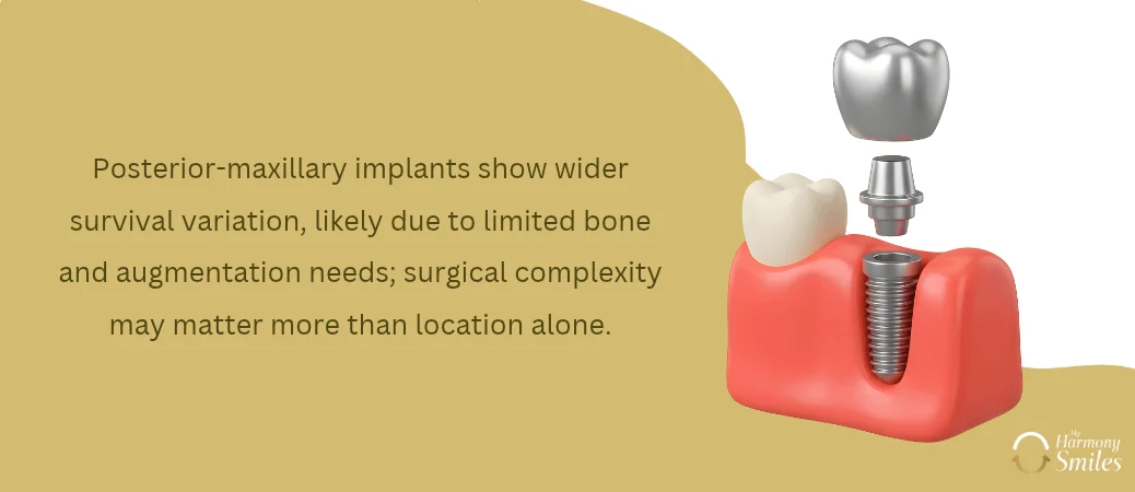
Among the four broad anatomical regions, the posterior maxilla has the widest survival range in the single-tooth systematic review, 85.7–100%.
This does not mean that an upper molar implant has an 85.7% expected survival rate. Rather, some protocols and populations within that heterogeneous literature produced lower estimates.
One explanation is that posterior-maxillary treatment may involve reduced residual bone height and sinus-related augmentation.
Patients exploring dental implants with bone loss may need additional evaluation to determine whether bone augmentation is appropriate.
A 2026 multicentre EHR study analysed 7,886 implants in 3,291 patients treated across nine university dental centres.
Simultaneous direct or indirect sinus augmentation was associated with more than twice the adjusted hazard of implant failure versus non-augmented controls, whereas staged sinus augmentation was not significantly different from controls.
The authors stressed that treatment complexity and timing appear important and that the retrospective coding-based design leaves potential confounding.
Similarly, the Minnesota study found lower survival in sinus-augmented groups and identified simultaneous ridge augmentation as a significant risk factor, whereas simply being in a posterior region was not independently significant.
This distinction is critical for research interpretation: a posterior location may sometimes act as a marker for a more difficult surgical environment rather than being the biological cause of failure itself.
This is an inference from the location and augmentation data rather than proof of causality.
Long-term survival means more than keeping the implant
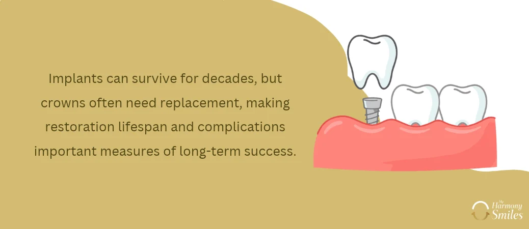
For single implants overall, the best older ≥10-year systematic review found an implant-level survival rate of about 95.0% among 527 implants, while survival of the original single crowns was lower, 89.5%.
The authors also cautioned that the number of genuinely long-term cases was small relative to worldwide implant use.
An extraordinary 2025 Swedish follow-up illustrates this distinction even more clearly. After 38–40 years, cumulative survival among the original single implants was 95.6%, but survival of the implant-supported crowns was only 60.9%; many crowns had been replaced, often for aesthetic reasons.
Only 18 implants in 13 patients were available at final follow-up, so this is evidence of what can occur over decades rather than a population forecast.
Consequently, research comparing “front and back implant success” should not use implant survival as the sole endpoint.
Long-term outcomes also involve implant maintenance, restoration complications, and the factors discussed in research on how long dental implants last.
The international 2023 ID-COSM consensus recommends also measuring peri-implant tissue health, surgical morbidity, adverse events, complication-free survival, prosthesis lifespan, and patient satisfaction/comfort.
Florida relevance and outlook beyond 2026
This search did not identify peer-reviewed, city-specific survival cohorts that permit separate single-tooth survival percentages for Ocala, Palm Harbor or Trinity.
University of Florida participates in contemporary implant research and the National Dental Practice-Based Research Network lists a Dental Implant Restoration Registry, but presently available evidence does not justify assigning different survival rates to these three Florida communities.
Accordingly, for research conducted in these markets, a defensible evidence hierarchy would use U.S. cohorts first, supplemented by international single-tooth site-specific studies where U.S. data are absent.
It would be misleading to advertise, for example, “98% front-tooth survival in Ocala” or a different percentage for Trinity without a local longitudinal cohort.
There is also no scientifically sound way, as of October 2026, to assign an invented “2030” or “2035” survival percentage.
The stronger post-2026 research direction is likely to be finer stratification: exact tooth number rather than simply anterior/posterior; early versus late failure; native versus augmented bone; immediate versus staged placement/loading; and implant survival separated from crown and complication-free survival.
The 2023 international core-outcome initiative specifically seeks more standardised reporting, while large EHR studies published in 2025–2026 demonstrate the feasibility of studying very large real-world populations.
Research conclusion: current evidence does not establish a universal front-versus-back winner. Anterior-maxillary single implants have the deepest location-specific evidence and generally very high survival; posterior mandibular implants also perform very well.
The posterior maxilla shows the greatest variability, apparently influenced partly by bone deficiency and augmentation requirements.
Anterior-mandibular evidence remains comparatively sparse and newer long-term data show that outcomes can be lower under particular protocols.
Overall, patient risk, local anatomy and treatment protocol provide a more scientifically useful explanation of survival than “front teeth versus back teeth” alone.
References
Implant survival in the anterior mandible: A retrospective cohort …
Outcome of Single Dental Implants Over 38-40 Years: A Long-Term Follow-Up Study – PubMed